Microscopy Image Competition
The inaugural Faculty of Medicine Microscopy Image Competition gives us a rare glimpse of the beautiful and complex world which can only be seen through a microscope.
Sixteen people from across the Faculty put their scientific and artistic skills to the test to bring us some truly stunning images.
The winners were selected by popular vote at the 2019 Faculty of Medicine Excellence Awards.
Winner
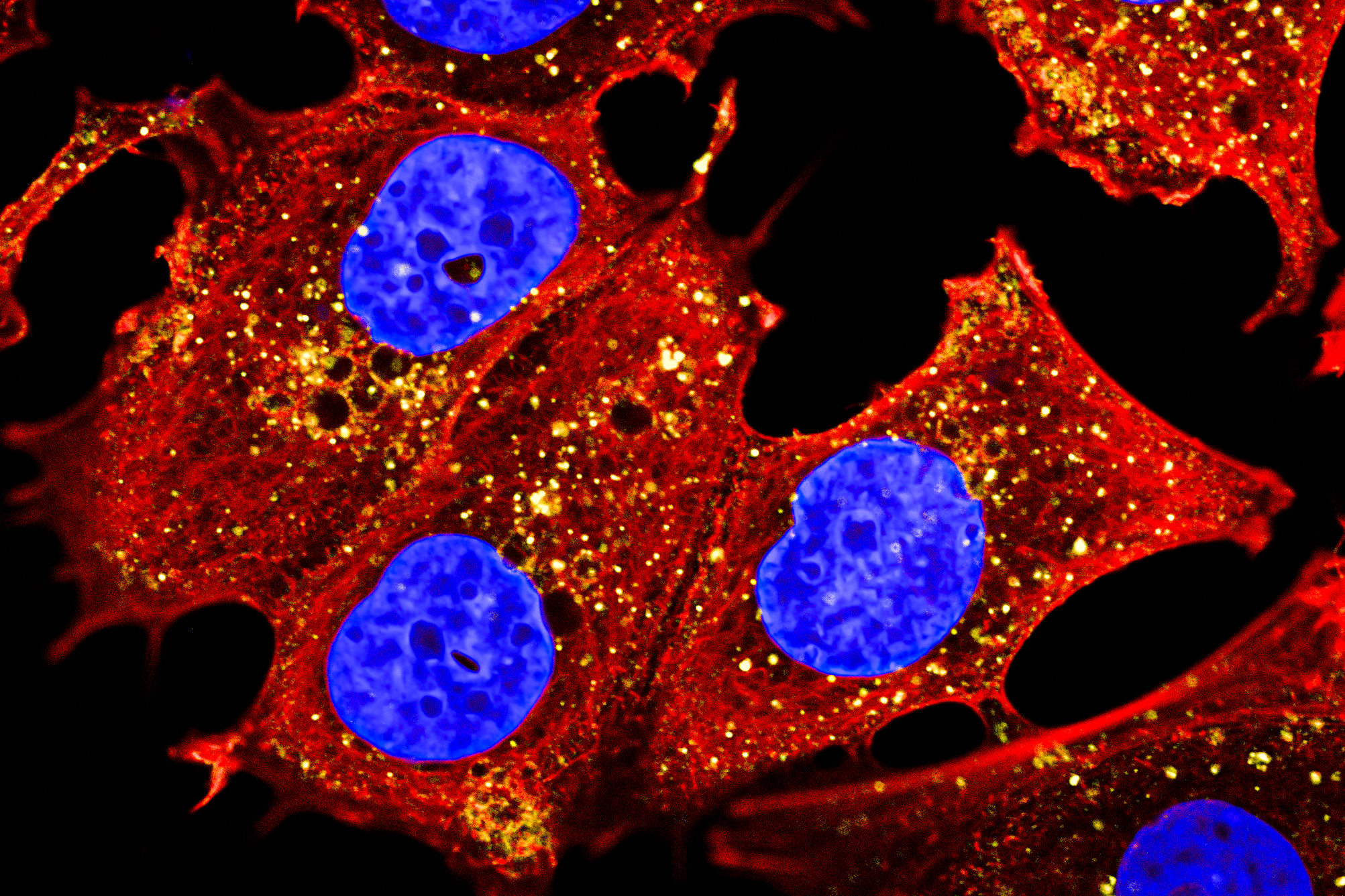
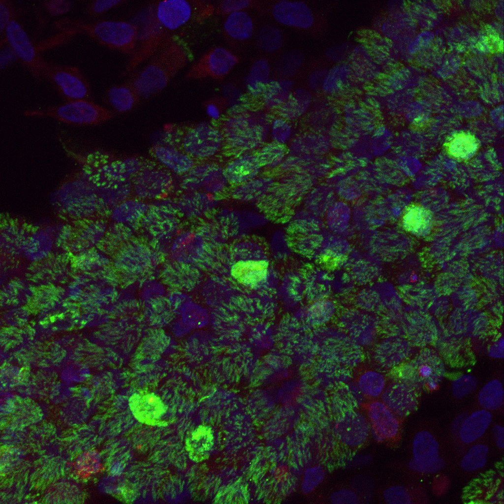
Internalisation of monoclonal antibodies in breast cancer cells
By Benedict Lum, Diamantina Institute
Instrument: Olympus FV3000 Point Scanning Confocal (TRI)
Anti-tumour effects of therapeutic monoclonal antibodies (mAbs) are highly dependent on its ability to mediate antibody dependent cell cytotoxicity (ADCC) carried out by the hosts’s immune effector cells. However, for ADCC to efficiently occur, mAb-target complexes must remain localised to the cell surface for immune effector engagement. Here, lumretuzumab is rapidly internalised into the cell, which prevents lumretuzumab from carrying out its function to mediate ADCC. This shows that having a mechanistic understanding of how targets are internalised during development of mAbs can affect the therapeutic efficacy of mAbs.
Second place
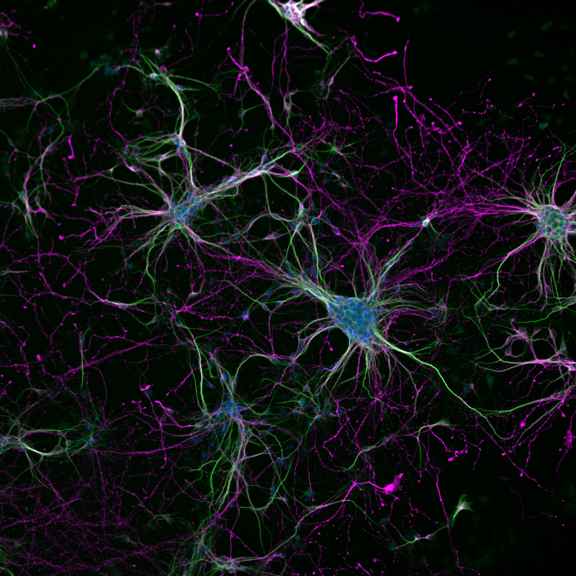
Making connections
By Maria Kasherman, School of Biomedical Science
Instrument: Diskovery Spinning Disk Confocal (SBMS)
These are cortical neurons taken from embryonic mice and grown in culture, stained with markers for axons and dendrites. The purple is Tau, a protein that is abundantly expressed in the axons of neurons. Therefore, misfolding and aggregates of Tau has been linked to Alzheimer’s disease.
The green shows Map2, a protein that is expressed in the dendrites of neurons and is crucial for interneuronal communication. The blue is DAPI, a commonly used marker for the cells’ nuclei. By culturing these neurons, we aim to replicate what might go wrong during neuronal development and investigate what mechanisms are affected.
Third place
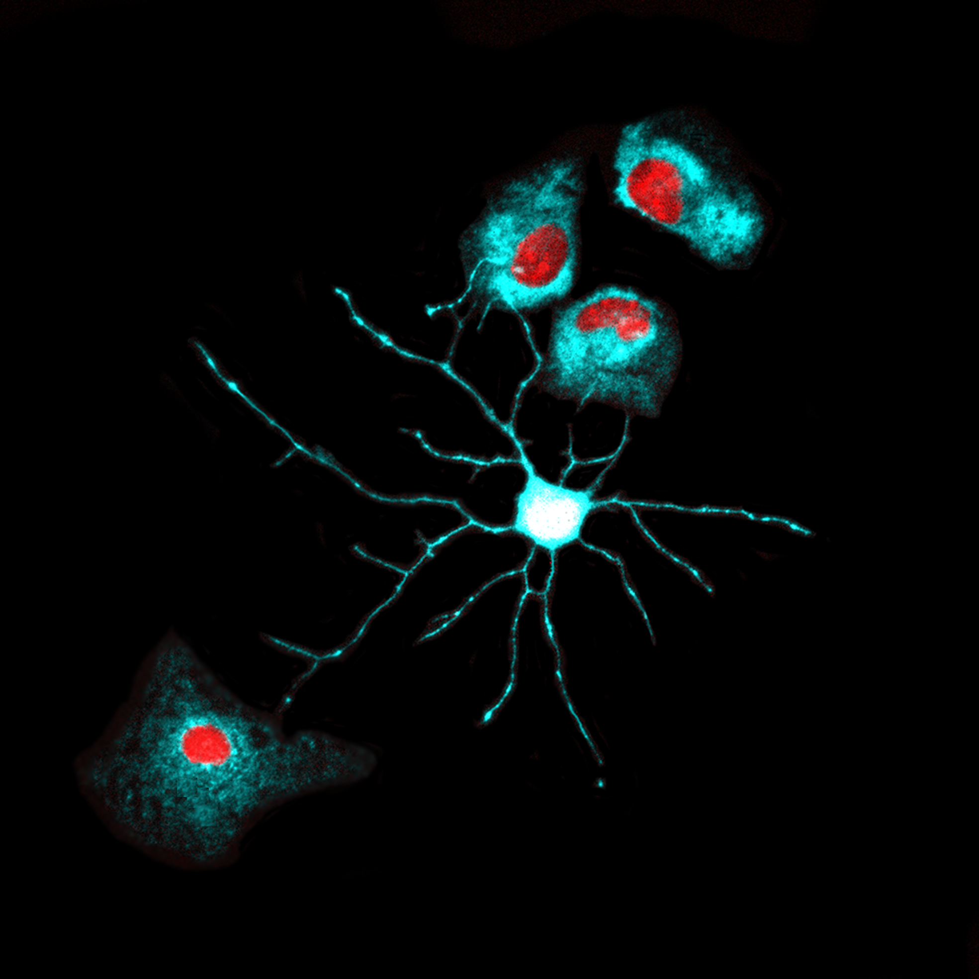
Shine Bright Like a Neuroblast
By Sabrina Oishi, School of Biomedical Sciences
Instrument: Diskovery Spinning Disk Confocal (SBMS)
The brain is comprised of over 100 billion cells, all originating from a small pool of neural stem cells. In vitro assays provide a powerful tool to examine the cellular and molecular mechanisms during the differentiation of neural stem cells. This image shows a single immature neuron (neuroblast) differentiated from primary mouse neural stem cells. The neuroblast’s dendritic processes are spread out, seeking to make connections with other cells. Eventually, neuroblasts will mature and integrate into a neural circuitry to create a functioning network. Thus, this image is a tiny snapshot of how billions of cells develop to make our complex brains.
Finalists
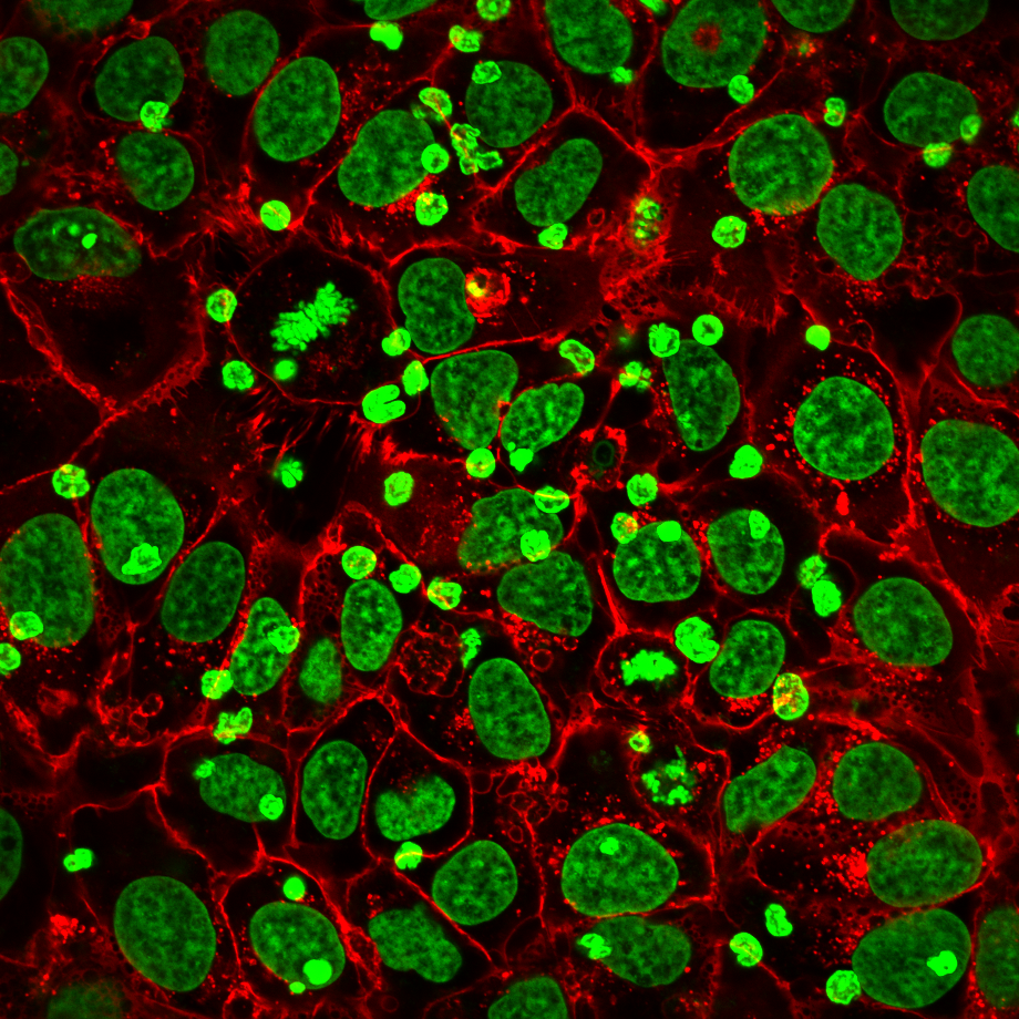
Immune Cells Attacking Cancer
By Blerida Banushi, Diamantina Institute
Instrument: Olympus FV3000 Point Scanning Confocal (TRI)
The humanised monoclonal antibody(red) binds to specific antigens on cancer cells (larger green nuclei) and this triggers the recruitment of immune cells (smaller green nuclei) that attack cancer cells.
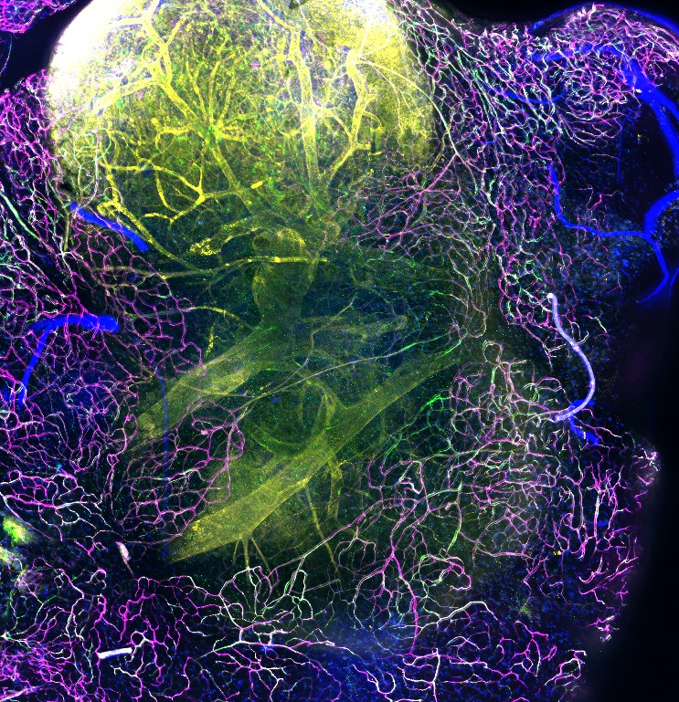
The Lymph Node Highway
By James Dight, Diamantina Institute
Instrument: Nikon Spinning Disk Confocal (TRI)
Lymph nodes are key centres for a functioning immune system. As they are highly vascularized, and act as reservoirs for unwanted goods, they commonly become colonised by cancerous cells which have typically disseminated from a primary location. We believe the endothelium in the vascular system, which runs adjacent to lymph nodes becomes activated by signals secreted by primary tumours. These signals prime secondary tissues to make them more accessible for colonisation by primary tumour cells. In our lab, we believe we have found new means of suppressing or turning off this activation event.
Galaxy in Brain
By Rui Li, School of Biomedical Sciences
Instrument: Leica SP8 Point Scanning Confocal (SBMS)
The exact function of MAP2 is unknown but MAPs may stabilize the microtubules against depolymerization. They also seem to have a stiffening effect on microtubules.
The products of similar genes in mouse are neuron-specific cytoskeletal proteins that are enriched in dendrites, implicating a role in determining and stabilizing dendritic shape during neuron development.
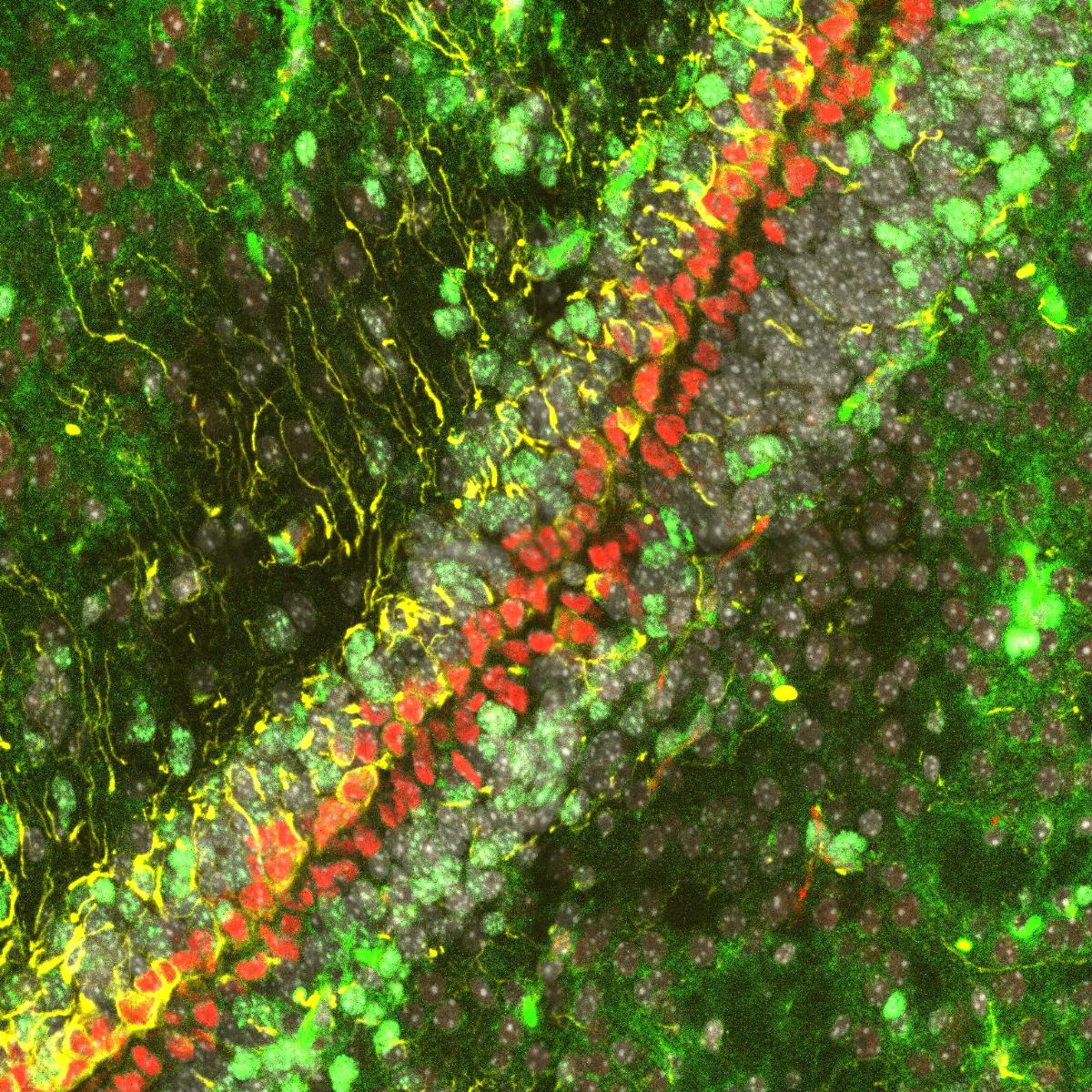
Roads
By Danyon Harkins, School of Biomedical Sciences
Instrument: Diskovery Spinning Disk Confocal (SBMS)
This image depicts a stain of the ventricles of a postnatal day 10 mouse. Ki67 (green) marks proliferating cells, while GFAP (yellow) marks glial cells and FOXJ1 (red) marks ependymal cells.
In wild type mice ependymal cells are non-proliferative following E18 and do not co-label for Ki67.
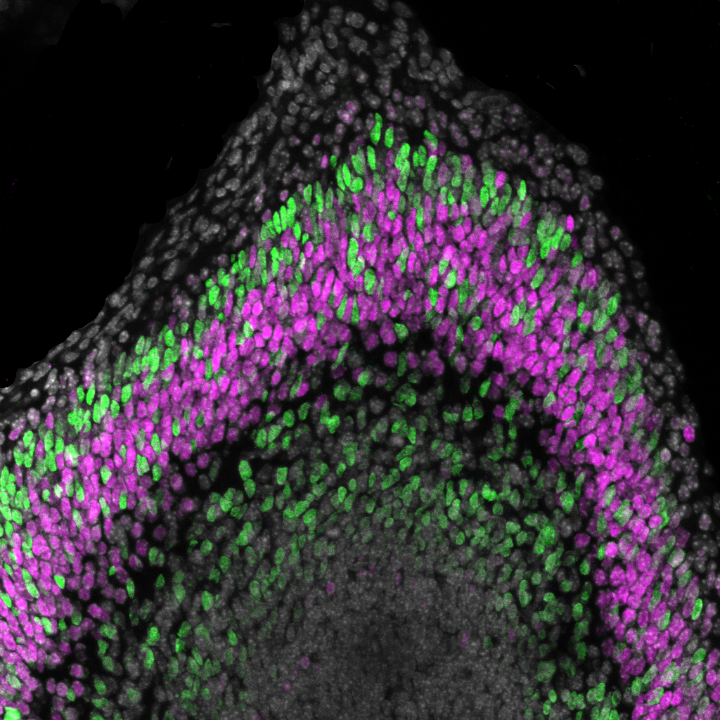
Neuronal Populations Immunostaining in Mouse Cortex
By Belal Shohayeb, School of Biomedical Science
Instrument: Leica SP8 Point Scanning Confocal (SBMS)
This staining was performed to investigate the neurogenesis process and neuronal migration in the mouse neocortex during development.
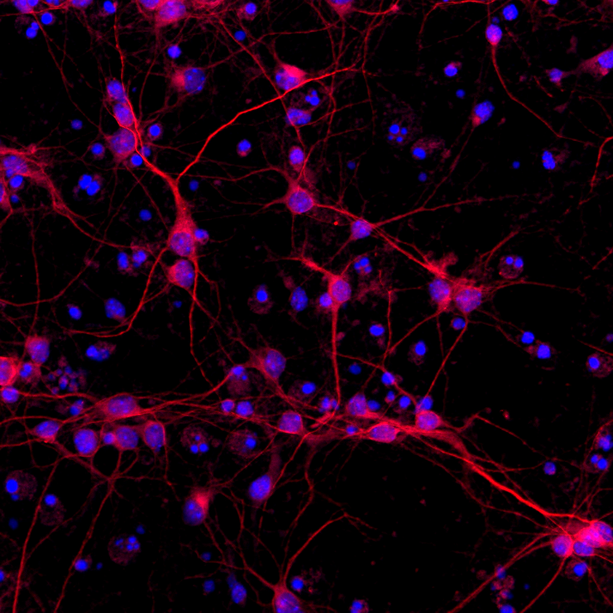
Networks in the Growth Restricted Brain
By Kirat Chand, Centre for Clinical Research
Instrument: Zeiss LSM710 Point Scanning Confocal (UQCCR)
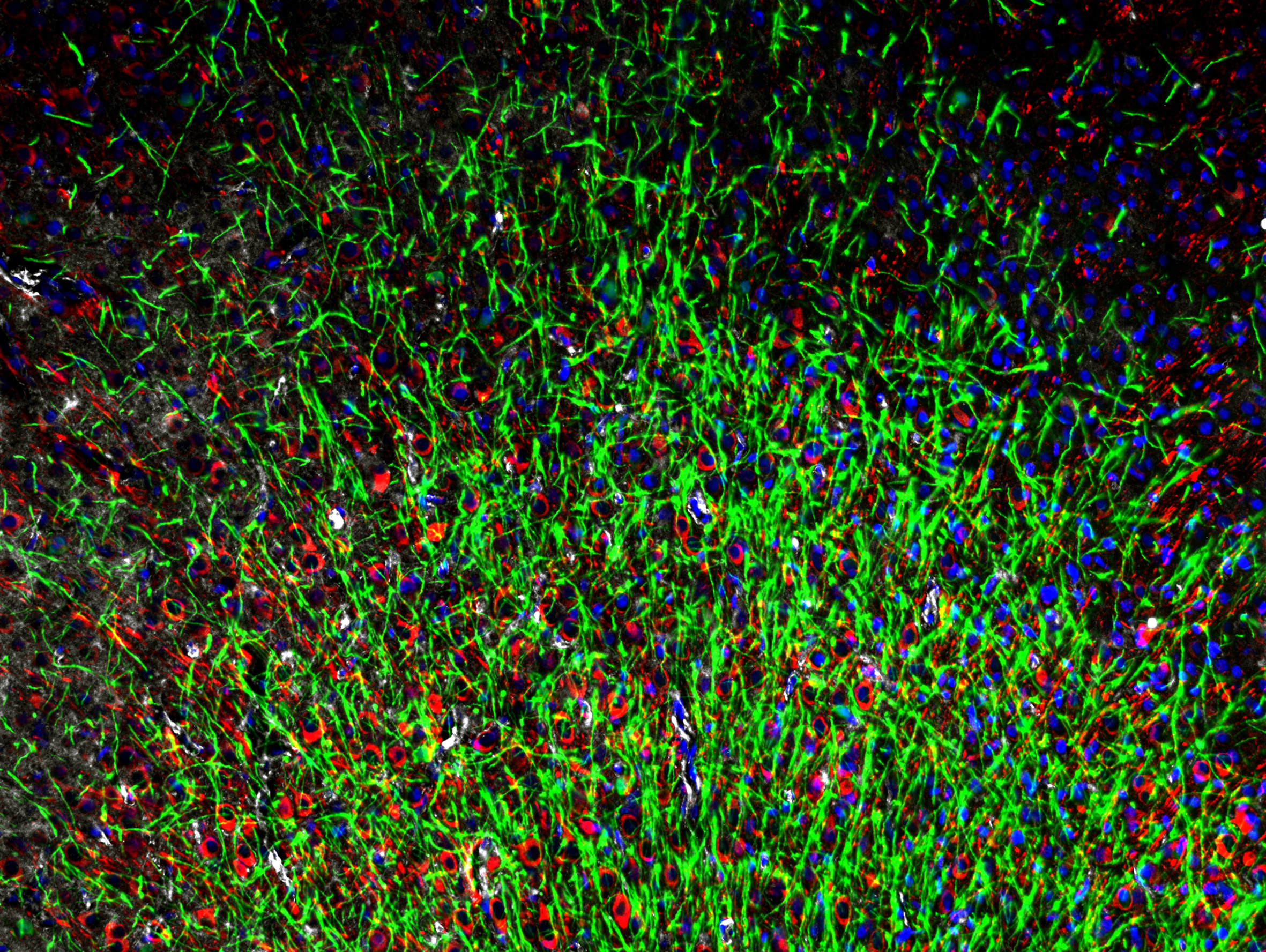
Neuronal networks are impacted by growth restriction in the neonate leading to alterations in brain health. MAP2 targeting dendrites of neurons (red) myelin basic protein - targeting neuronal myelin (green) and GFAP targeting astrocytes (white) shows the networks that are forged in the developing brain and the complicated interaction that allows optimal function. These components are often disrupted as a result of growth restriction and are a possible target for treatments.
HER2-HER3 Interactions in Brain Metastases
By Malcolm Lim, Centre for Clinical Research
Instrument: Zeiss Axio M1 Imaging Microscope (UQCCR)
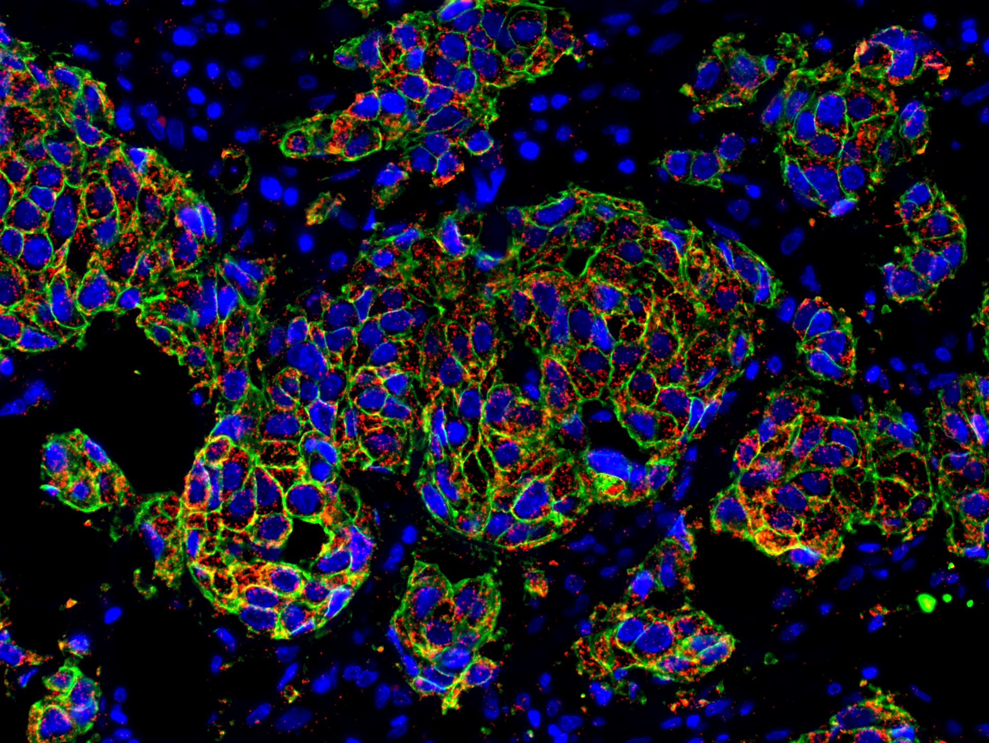
Tumours with HER2-HER3 dimers are more resistant to HER2-targeted treatments. This is because when the HER2 and HER3 proteins interact, their dimer-complex triggers an intense proliferation and survival signal in tumour cells. Blocking this “escape” mechanism is essential but evidence is lacking. Common immunofluorescent technique is limited to imaging colocalization of proteins. Through the use of an in situ proximity ligation assay kit, visualisation and quantification of HER2/3 dimers was made possible. My research demonstrated that HER2/3 dimers are indeed prevalent in brain metastases, and that combination targeting of both HER2 and HER3 is a potential therapeutic strategy. This image depicts a section of breast cancer brain metastasis. Cell nuclei are denoted in blue, the HER2 receptor protein in green and each red dot represents a HER2/3 dimer.
Skin Deep
By Robert Ju, Diamantina Institute
Instrument: Nikon Spinning Disk Confocal (TRI)
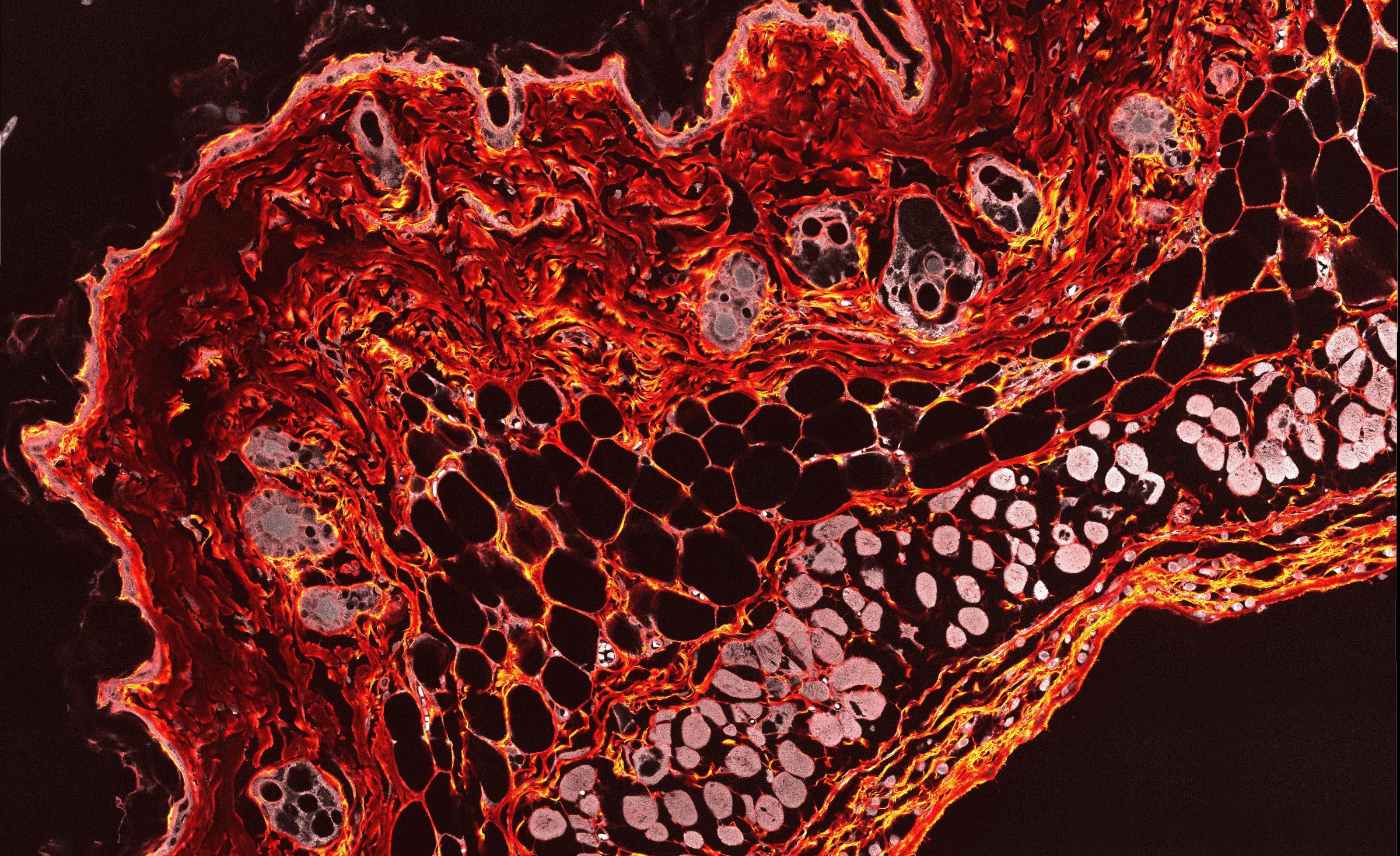
The skin is our first line of defence, forming a physical barrier that prevents infection.
Our skin is remarkable as it is formed by the multiple intricate cell types that co-ordinate to form multiple complex layers.
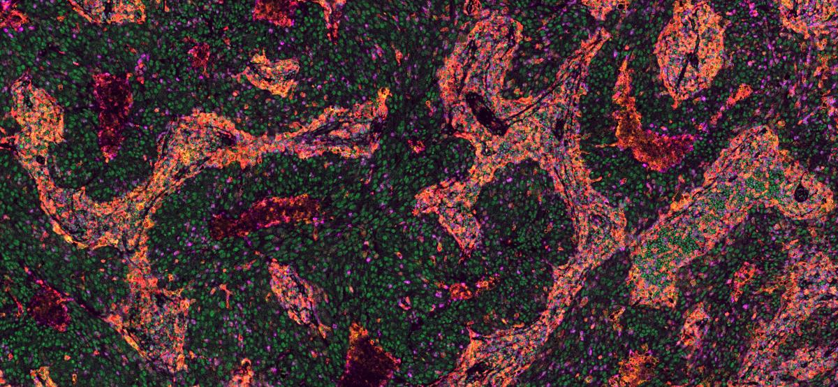
The Complex Microenvironment of Human Breast Cancer
By Priyakshi Kalita-de Croft, Centre for Clinical Research
Instrument: Zeiss Axio M1 Imaging Microscope (UQCCR)
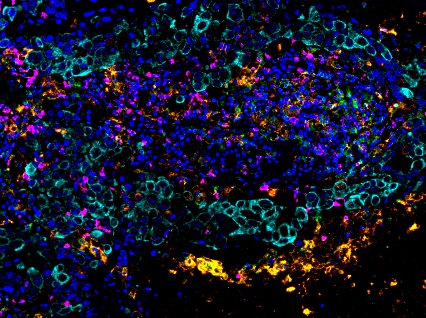
This image depicts the complex micro-environment of a human breast cancer.
An army of immune cells (dark blue, green, magenta and orange) are invading in and around the cancer cells (cyan) trying to control and destroy them.
Entries
Primary Human Nasal Epithelial Cells
By Ayaho Yamamoto, Centre for Children's Health Research
Instrument: ZEISS LSM710 (UQCCR)
When the ciliated cells appear, it means the primary human airway epithelial cells are well-differentiated at air-liquid interface. Therefore, staining ciliated cells to show the differentiation.
Compared with the control, this image shows that mitochondrial reactive oxygen species (mtROS) production increased after exposing to oxidative stress. Increase in mtROS can causes mitochondrial dysfunction and leads to respiratory diseases such as chronic obstructive pulmonary disease. This image can also be compared with cells pretreated with anti-oxidant. The result showed that cells pretreated with anti-oxidant decrease in mtROS production.
Blue: nuclei, Green: ciliated cells, Red: mtROS
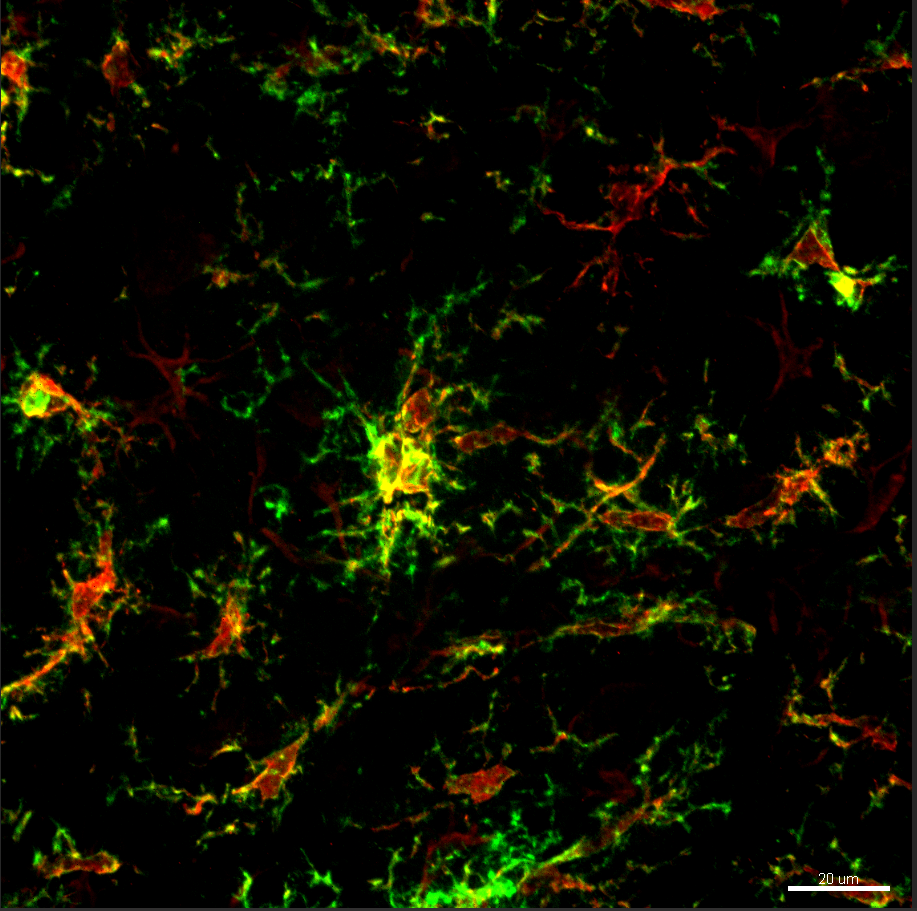
A Microglial Menagerie
By Sandra Parker, School of Biomedical Sciences
Instrument: Diskcovery spinning disk microscope (SBMS)
Increasing neuroinflammation in this mouse model of ALS is associated with increases in motor neuron cell death, leading to paralysis of the hind limbs in these animals.
Characterising this neuroinflammation is the first step in developing novel therapies for ALS, as slowing microglial infiltration and activation may represent a strategy to limit motor neuron death in these animals.
Image of microglial markers (IBA-1 in red, CD11B in green) in the spinal cord of a mouse model of amyotrophic lateral sclerosis. This image shows activated microglia in the lumbar spinal cord.
Melanoma immune heterogeneity
By Vanessa Bonazzi, Diamantina Institute
Instrument: Zeiss AxioScan Z1 Scanner (SBMS)
Melanoma tumours are highly heterogenous. The immune microenvironment in the tumour is a key factor determining whether a patient will respond to therapy.
This multiplex labelling technique highlights the heterogeneity of the immune cells infiltrating the melanoma tumour tissue. Each colour highlights a different type of immune cell with specific roles relating to immune response.
Inflammation in hypoxic-ischemic neonates
By Lara Jones, Centre for Clinical Research
Instrument: Zeiss Axio microscope (UQCCR)
Hypoxic-ischemic neonatal piglet model used to highlight inflammation is a driving force of injury in the brain. Image shows th staining of astrocytes (green) and activated microglia (red) in the injured brain, inflammation is increased in the injured brain - seen by the high numbers of activated microglia and astrocytes stained in the cortex of this animal (dapi stain indicates cell nucleus).
