Looking down the microscope collection

Historical perspectives
According to various ancient texts, precursor microscopes appear to have been developed in various forms by the Chinese some 4000 years ago (the water microscope) and with it they could achieve magnification of 150 fold. Aristotle described the working of a basic microscope in some detail and the Greeks made good use of early curved lenses. However, there are virtually no references to the science of optics until the late 13th and early 14th centuries when the invention & use of spectacles was described in Italy by Amati, Rivalti and others. At this time, the development of telescopes had begun and was described by the Englishman Roger Bacon.
Progressive improvement in the quality and sophistication of lenses facilitated the further development of microscopes and some of the noted scientists involved included Galileo Galilei and Isaac Newton. There has been much debate over who invented the microscope and to some extent this is related to the classification as either “simple” (single lens) or “compound” (two lenses) microscopes. Zaccharias Janssen, a Dutch spectacle maker, described his compound microscope circa 1590 and some 70 years later Robert Hooke, the English polymath, produced his own microscope.
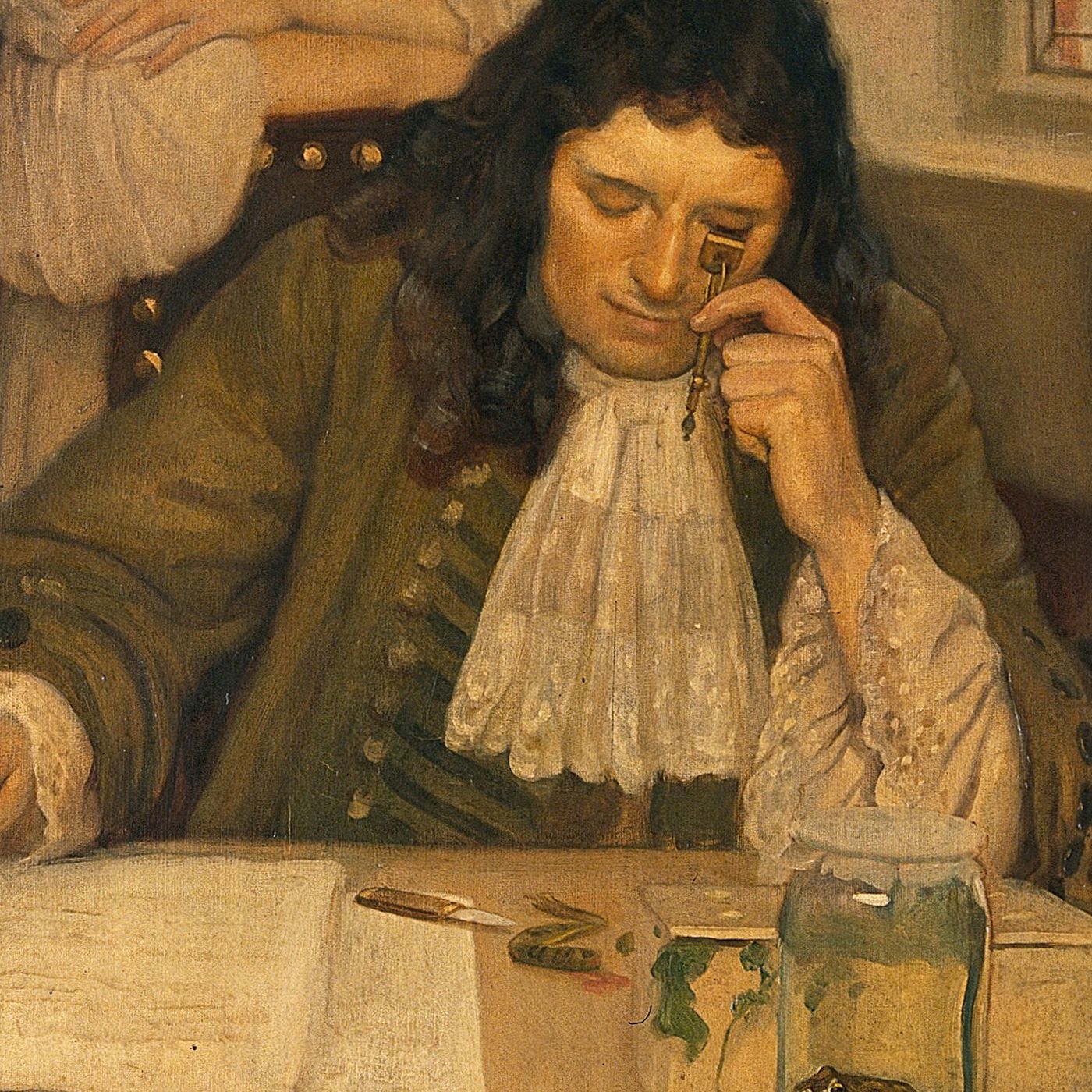
At about the same time (1665), the Dutch cloth merchant and part time scientist, Anthony Van Leeuwenhoek produced his simple single lens microscope. The superb lens that he had made provided better magnification and clarity than any of the other existing microscopes and he has been called the “father of the microscope”. He famously became the first person to describe bacteria as well as human and animal red cells.
It was in the 1730s that Charles Hall devised an achromatic lens system which overcame the problem of blurring and rainbow effect (spheroidal aberration) that occurred at high levels of magnification. This lens system was further improved by Joseph Lister in 1830. The quality of microscope lenses continued to improve and in 1860 Ernst Abbe, a professor of physics and mathematics and a partner in the Zeiss Microscope company, devised a framework for modern optics that eliminated problems between the magnification and resolution of lens systems. In 1869 he produced the Abbe Condenser which radically improved the quality of light being passed from its source (then usually a purpose-built spirit or kerosene lantern) to the microscope lens system. It was not until the development of the Edison incandescent light bulb in the 1890s that a better source of illumination was introduced. It was a further 40-50 years before “on-board” electric light bulbs, condensers and filters became the norm. Other features such as dark ground illumination, polarising light lenses and fluorescence microscopy soon followed. Binocular microscopes were in existence as far back as the 1860s but only became common place and virtually universal in the 1960s. Other types of optical microscopes with specific features have been developed for a variety of applications and these include stereoscopic, phase contrast and differential interfering contrast instruments. Highly specialized research instruments developed during the twentieth century include the ultramicroscope (Richard Zsigmondy 1903), phase contrast microscope for colourless & transparent biological materials (Fritz Zernike 1932), the electron microscope (Ernst Ruska 1938) and the scanning tunnelling electron microscope (Gerd Binning and Heinrich Rohrer 1981). The inventors of all four of these instruments were Nobel Prize recipients.
The contribution of microscopy to medicine has been immense particularly in the development of our understanding of disease processes, their causes and therapy. It now seems surprising that prior to the 1840s microscopes were used primarily for non-medical purposes such as entomology and botany. In the 1840s several physicians described patients with a variety of symptoms such as enlargement of the liver, spleen and lymph nodes and “suppuration of the blood”. John Hughes Bennet of Edinburgh, in reporting his patient, recognized that the high white cell count was not due to inflammation. In the same year, Rudolf Virchow in Berlin, reported a similar patient and in 1846 Henry Fuller of London reported other cases. These patients had somewhat different clinical findings and varied course of disease which probably reflected the different types of leukaemia involved. It was not until the 1850s that microscopy became an integral component in medical diagnosis. This was largely due to the work the Prussian physician Rudolf Virchow known as the “Father of Pathology”. He was instrumental in correlating anatomical changes seen in various disease states and with the patient’s symptoms. He developed an interest in microscopy and cellular pathology and his first scientific paper published was a description of “leukaemia” – a term that he invented (this is disputed because “leukocythemia” had been used previously). Microscopy was soon embraced by other specialties within pathology especially haematology and bacteriology. This was facilitated by the introduction of specimen staining techniques which assisted differentiation of the various cell types in blood and other specimens. The stains were initially introduced by Paul Ehrlich and were derived from aniline dyes used in the textile mills of Silesia and they were subsequently improved by the Russian physician Dmitri Romanowsky. Further modifications by Jenner, Giemsa, Leishman, Grunwald , Wright & others improved the stains further and they are known as the Romanowsky group of stains and they are routinely used in haematology laboratories today. In haematology the stains facilitated the diagnosis of various anaemias, leukaemias and parasitic infestations such as malaria. Ehrlich’s acid- fast stain was modified by his countrymen Franz Ziehl and Friedrich Neelsen and it enabled the tuberculosis bacillus to be identified. Hans Gram, a Dane developed the stain that bears his name and it remains a primary tool in the identification of bacteria to this day. Paul Ehrlich was awarded the Nobel Prize in 1908 for his research into the treatment of syphilis with arsenical compounds.
Significant advances in laboratory technology and instrumentation since the 1960s, particularly in haematology, has seen the replacement of much laborious manual testing techniques (such as haemoglobin measurement and blood cell counts) by the introduction of automated specimen processing. In 1979, the “Hematrak” (Geometric Data Corporation), an automated differential white cell system was introduced. It was in essence, a high quality Zeiss optical microscope with a motorized stage integrated to a pattern recognition computer and it was capable of processing 100 blood films per hour. This revolutionized work flow in large diagnostic laboratories in North America and several of the instruments were installed this Australia. However, this technology was subsequently replaced by newer systems where all the components of a full blood count are measured and analysed in fluid blood in a continuous flow system. Light microscopy has not entirely been replaced and is still essential when the analyser detects a significant abnormality in the blood specimen or the patient’s clinical history warrants manual examination by a skilled scientist or pathologist. Likewise, there have been significant ancillary advances in microbiology and histopathology over the last few decades, including immuno-peroxidase staining and molecular studies, but light microscopy will remain an essential tool of diagnosis in these disciplines for the foreseeable future.
The Marks Hirschfeld Collection
The MHM microscope collection comprises some 35 catalogued instruments. A further 5 uncatalogued duplicated instruments (student models) are the residue of the class instruments which were used for teaching histopathology, microbiology and parasitology in the Mayne Medical School. Approximately one half of the instruments are constructed of brass and are of European manufacture – predominantly German (Leitz Wetzlar, Zeiss Jena). Of the remainder, Japanese instruments predominate (Olympus, Tiyoda, Yashima). No manufacturer’s mark is present on 5 of the instruments. It is difficult to accurately establish the age of many of these instruments and relevant records, provenance etc. were unfortunately not a priority in the early days of the museum. Only two instruments have the date of manufacture marked – both made by the American company Bausch & Lomb some 20 years apart (1894 and 1915 respectively). Some instruments do have serial or model numbers engraved on them but this has not proven particularly helpful with older instruments where manufacturers’ records may be unavailable and indeed some of the companies no longer exist.
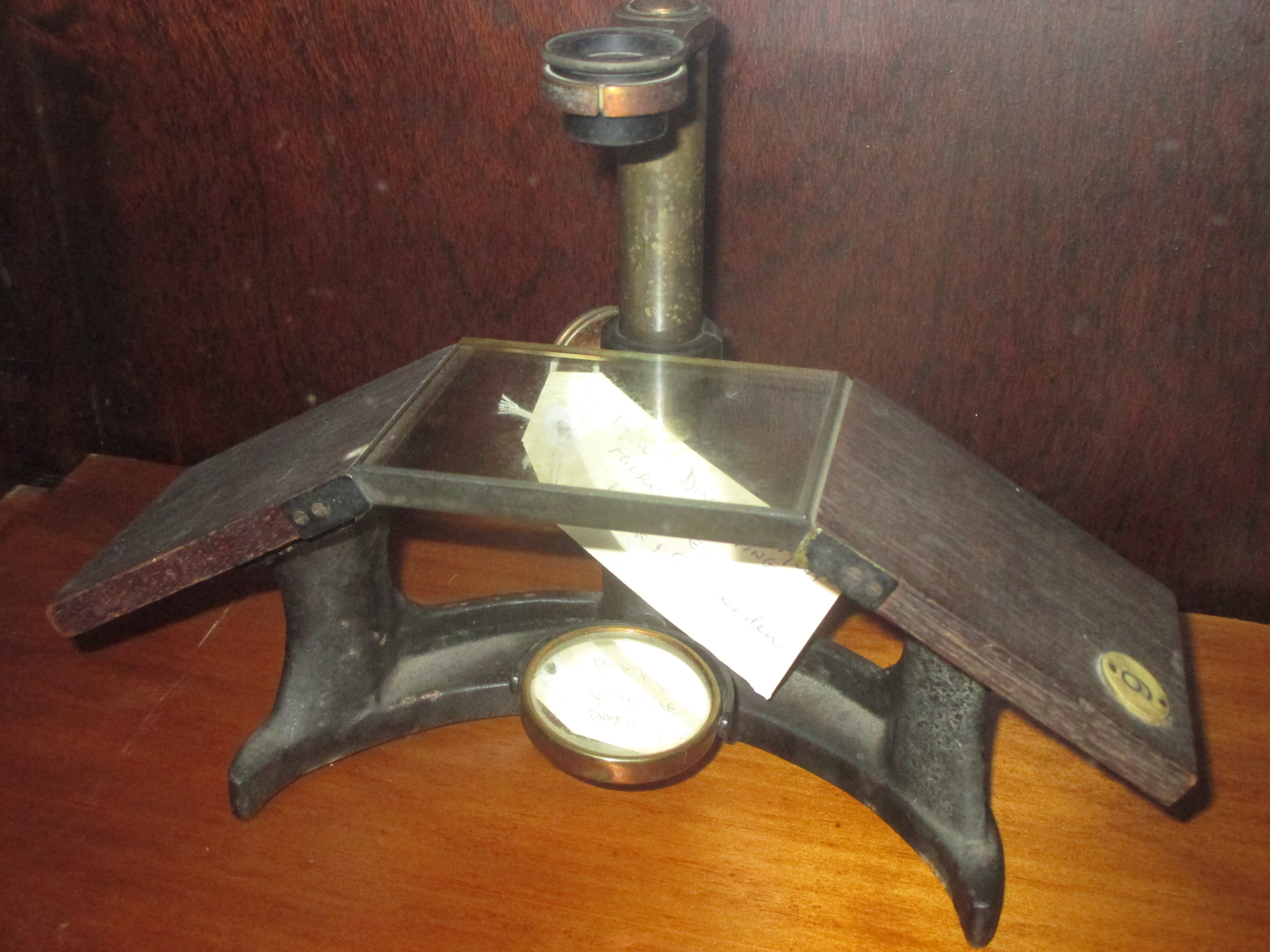
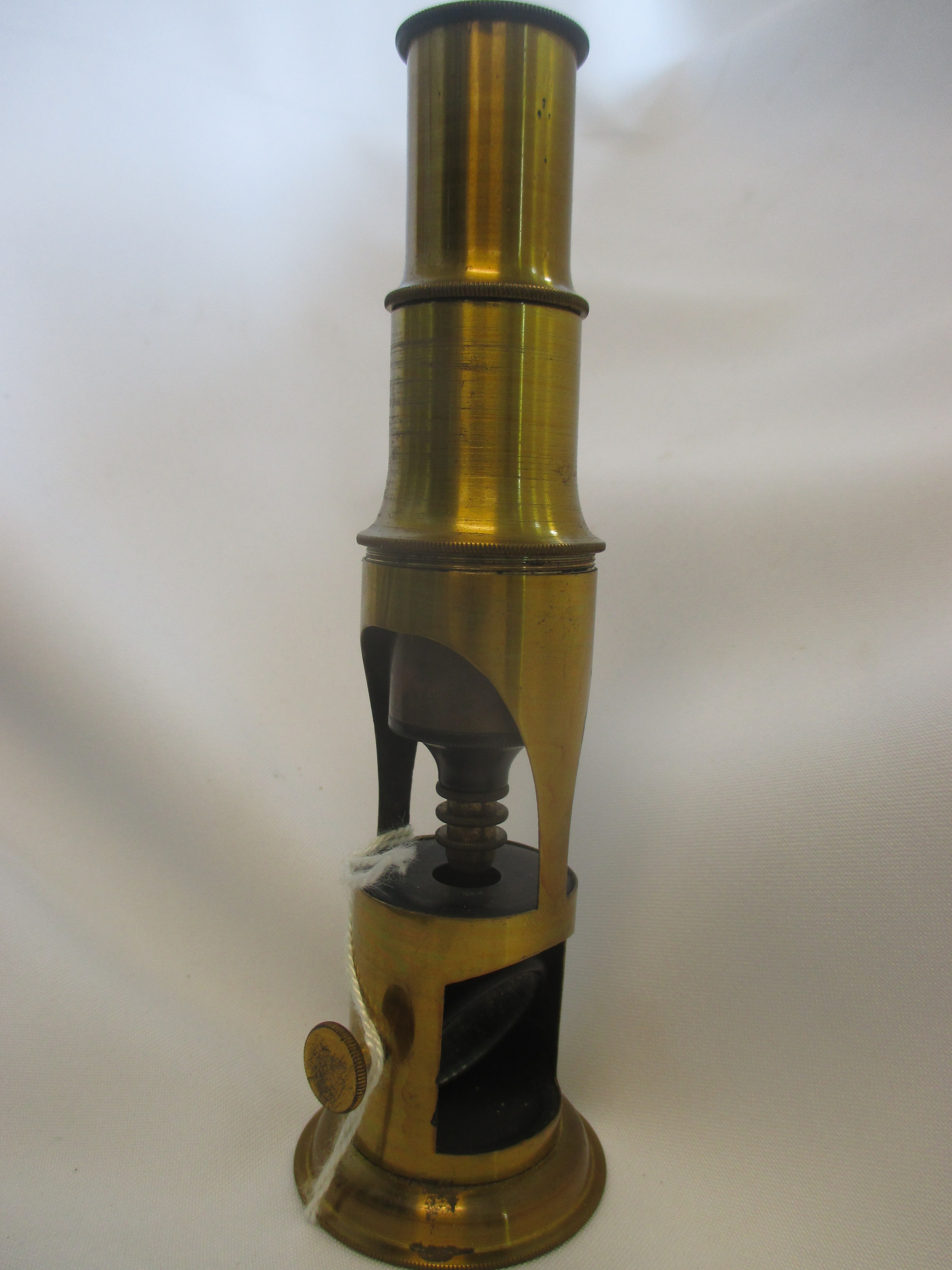
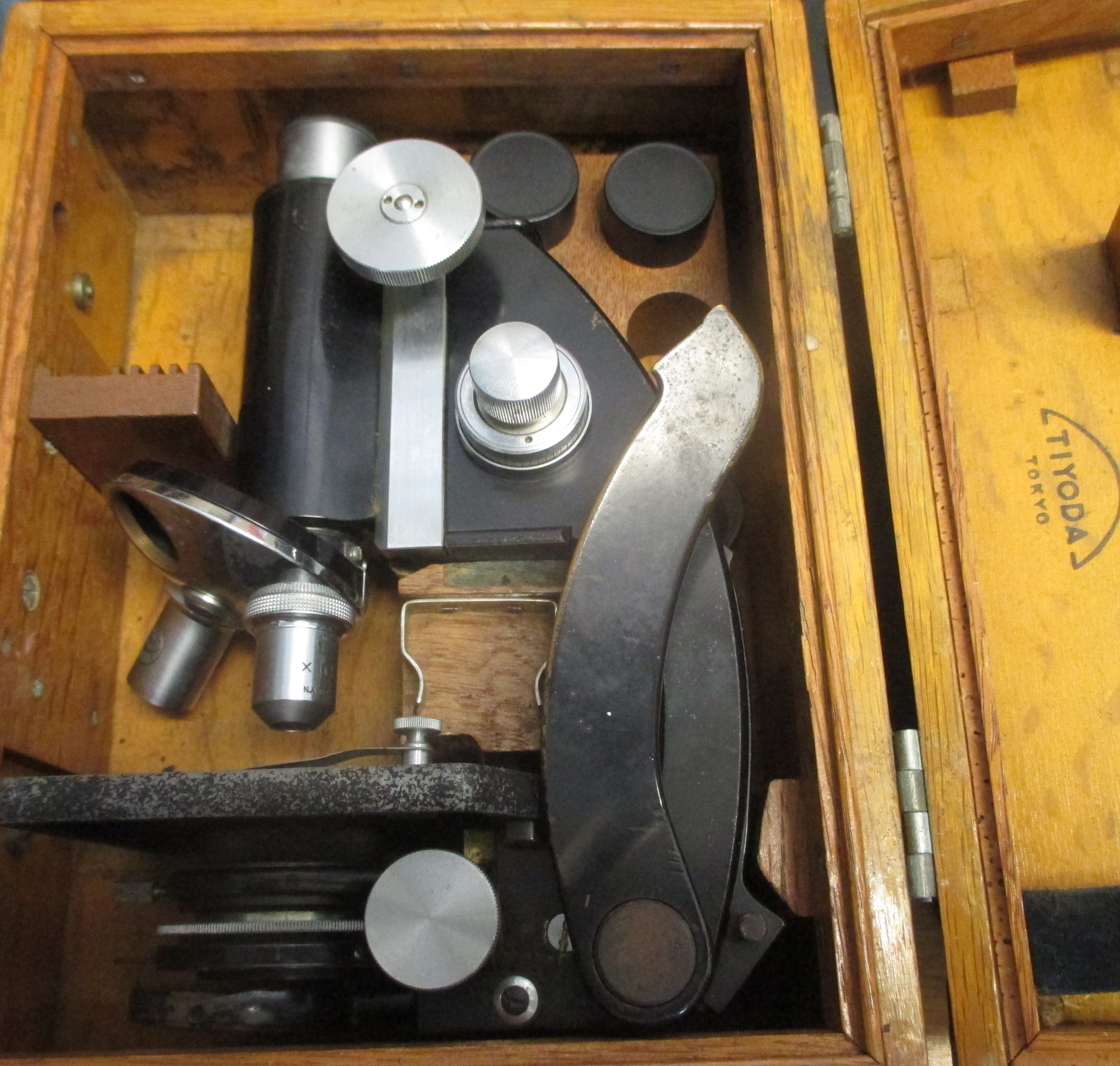
However, it is possible to make a few generalizations. Production of brass microscopes essentially ceased at the onset of the First World War when undoubtedly other uses for brass took priority. Virtually all microscopes made after that date were constructed in black enamelled iron until the 1960s when other colours such as grey and beige were and are being used. Another useful fact is that the under-stage light condenser devised by Ernst Abbe was produced from 1869 onwards. Thus, it is reasonable to assume that our brass microscopes which do not have an Abbe type condenser are likely to have been produced prior to1869 and are more than 150 years old. There are two quite rare instruments in the collection, the first is a small brass drum (pocket size) microscope circa 1840 and the second is a brass instrument with an attached spirit burner light source circa 1850 made by Allen and Hanbury. Other instruments of interest include an early dissecting microscope Circa 1870, a brass microscope Circa 1870 which belonged to Dr Jefferis Turner (first full-time doctor appointed to the Brisbane Children’s Hospital), A Zeiss steel & brass instrument circa 1930 which belonged to Dr Josephine Mackerras (medical researcher & grand-daughter of Dr Joseph Bancroft), and finally a small Tiyoda Japanese folding field microscope Circa 1942.
The Marks-Hirschfeld Museum of Medical History aims to celebrate Queensland’s medical history by telling the stories of its people, events, objects, scandals and triumphs. We welcome all stories with a medical history aspect. Get in touch with us at medmuseum@uq.edu.au.