“A new kind of Rays” comes to Queensland

Whilst investigating the effects of passing an electrical current through a vacuum tube on November 8th 1895 Professor W.C Roentgen observed that emissions from the tube caused a nearby screen to fluoresce. After further experiments he then hypothesized that the tube was emitting a new kind of ray, invisible to the eye, which could penetrate solid objects. On 28th December 1895 Roentgen delivered a paper “On a new kind of Rays” to the secretary of the Wurzburg Medical Society. This paper was subsequently published in the journal Nature on 23rd January 1896. The discovery was reported in the British Medical Journal on the 18th January 1896. Two weeks later On the 31st January 1896, the discovery was reported in the Brisbane Courier.
The apparatus required to produce the so-called X-rays was relatively simple. There were three components. Firstly, a power source which at that time consisted of an early electric primary cell known as a Groves Cell. Three of these cells are then connected to an Induction Coil. This is a device, which converts the low-pressure electric energy into very high-pressure electric energy in the form of a pulsating current. The third component is a glass vacuum tube. Through the walls of the tube are sealed 2 electrodes. X-rays are generated when a beam of high energy electrons emanating from the Cathode hits a metal target (the Anode). The image of the object of interest could then be captured either on photographic paper, or on a fluorescent screen.
The first documented instance of a successful radiograph in Australia was published in THE ARGUS on 4th March 1896 and referred to the work of Thomas Ranken Lyle a physicist and Professor of Natural Philosophy in the University of Melbourne. Although Lyle; by way of his profession had ready access to all three components listed above; he was also a skilled glassblower; and was able to make his own glass vacuum tube! The report detailed the successful procedure carried out on the previous day, which resulted in the image of the left foot of his colleague Orme Masson, Professor of Chemistry at the University of Melbourne.
Experiments with X-rays were first carried out in Brisbane on 16th July 1896 by Mr J.W. Sutton in his laboratory at 29 Eagle Street. The laboratory was directly across the river from Mr Sutton’s foundry at Kangaroo Point. Mr Sutton was well known in Brisbane as an ardent experimenter in science and a clever amateur photographer. The experiments were made possible by Mr Sutton’s acquisition of a coveted Crookes vacuum tube. They were carried out in the presence of Drs Love, Lyons, Rendle, Taylor and Wheeler as well as Mr Campbell, president of the Brisbane Photographers’ Society. On this occasion the images of various objects were displayed on a fluorescent screen.
There was a subsequent presentation to the Royal Society of Queensland on 8th August which was also attended by two surgeons; Dr Wilton Love and Dr David Hardie who were so impressed that they subsequently became the first doctors to use X-rays for clinical purposes in Queensland.
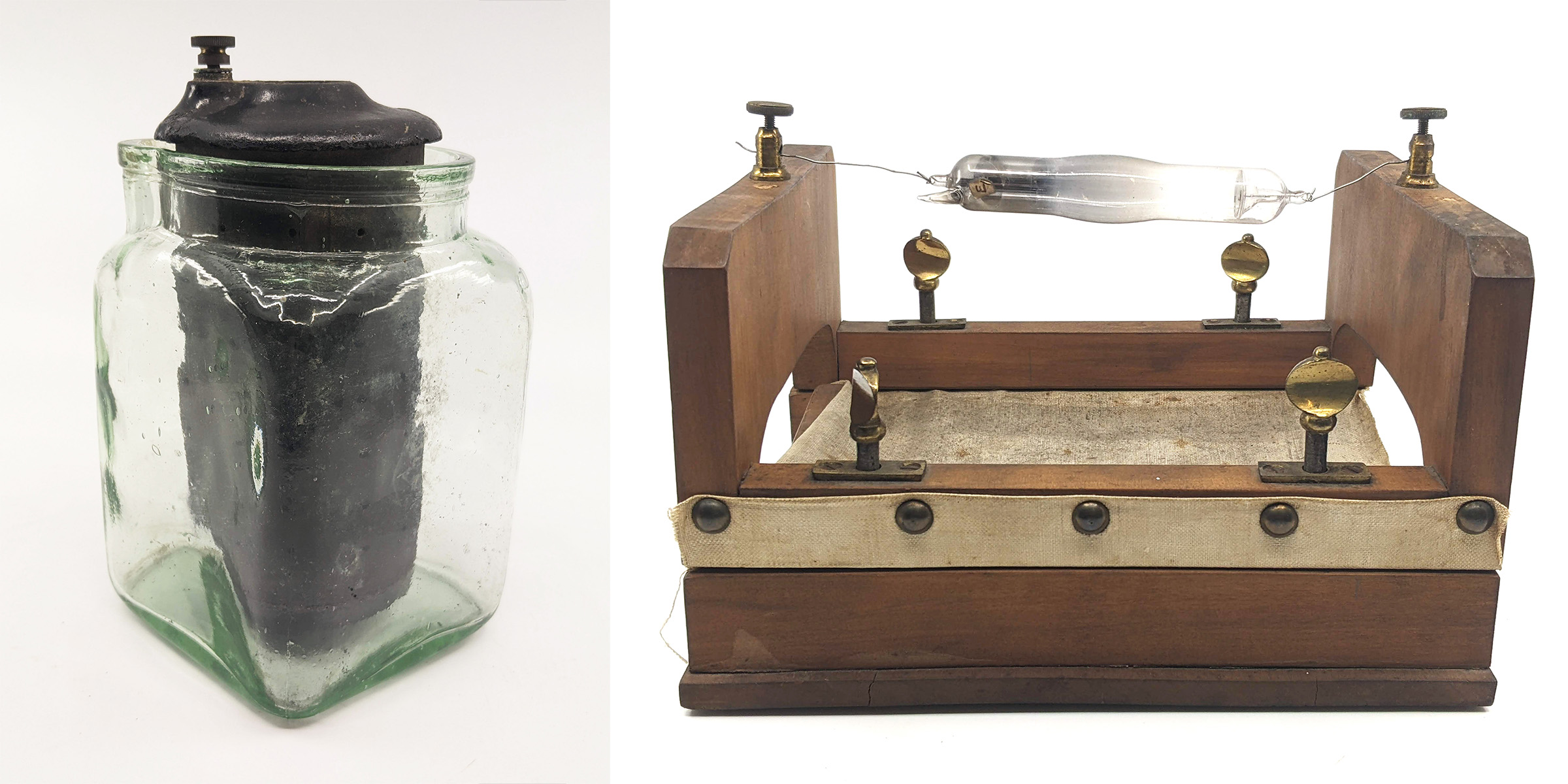
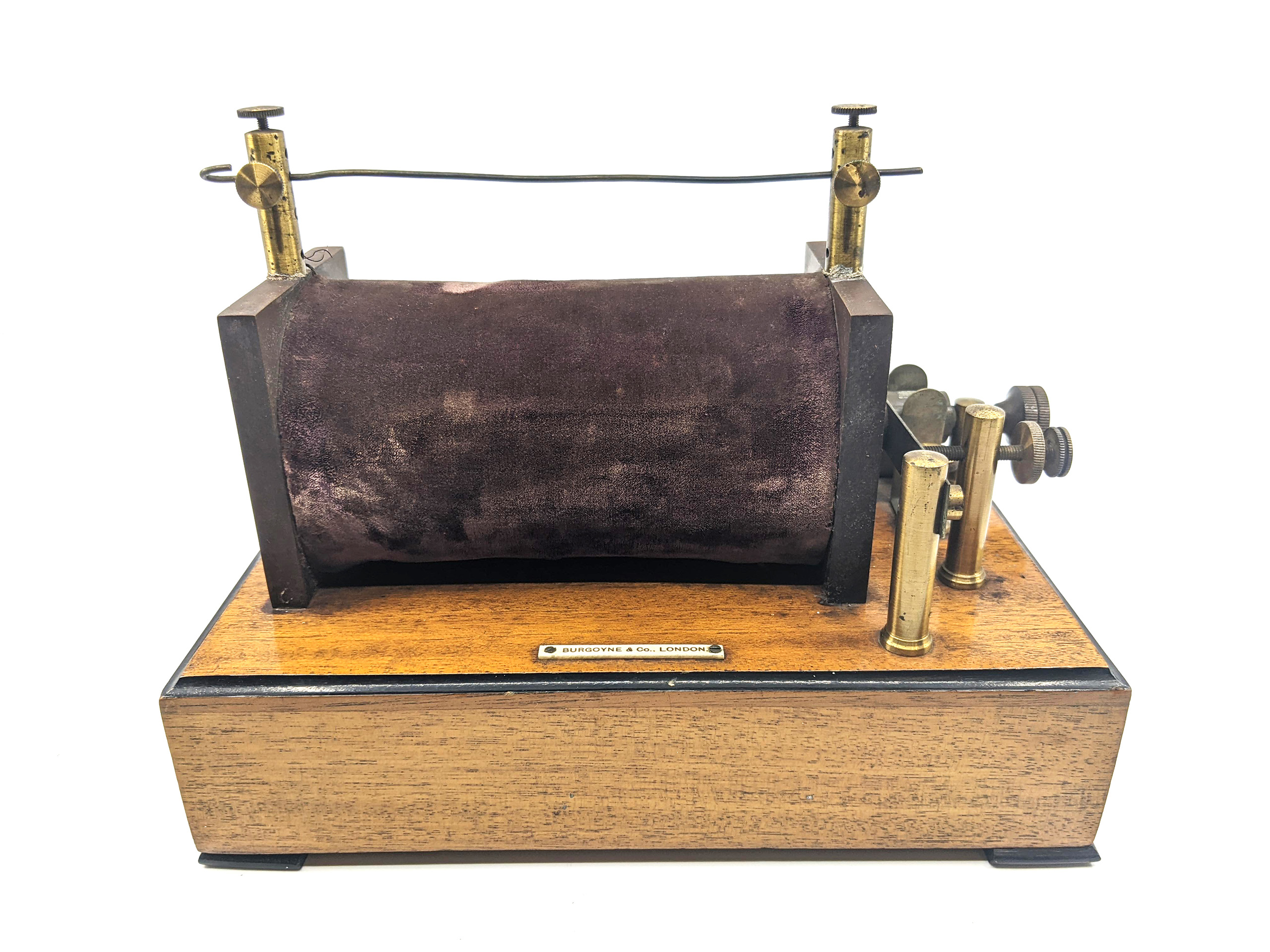
On the 11th June 1896 an X-ray apparatus comprising a vacuum tube (125mmx75mm); an induction coil; and 3 batteries; which was purpose built by Brady and Martin Ltd, Manufacturing Chemists and Surgical Instrument makers of Newcastle-upon Tyne; was forwarded to Brisbane by Burgoyne & Burbridges Manufacturing Chemists and Photographic Exporters of London. This was accompanied by a typed set of instructions, which unfortunately did not include the address of the consignee. The vacuum tube is mounted upon a wooden frame measuring 200 x 230 x 140mm. Below the tube is mounted an adjustable canvas sling which can accommodate an open hand. Below the sling is a sliding tray for a photographic plate.
This is not the apparatus that Mr Sutton utilised in his initial demonstration as the vacuum tube is not either a conventional Crookes or alternately a Hittorf Tube. It seems likely that the apparatus in question was consigned from London to either Dr Wilton Love or to Dr David Hardie, as these two gentlemen would have both been acutely aware of Roentgen’s exciting discovery and eager to acquire an X-ray apparatus.
The report of the Royal Society meeting on 8th August describes that again on that occasion a fluorescent screen was utilised which facilitated the display of the objects examined to the audience. It is noted that the “observers were able to see at once the bones of the hand, coins in a book etc”
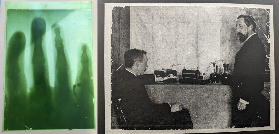
This first array of X-ray apparatus together with the sheet of instructions eventually came into the hands of Mr Fred L’Estrange. He was a senior executive in the Southern Electric Light Company and subsequently gave the apparatus to the collection of historical artefacts at the UQ Medical School; now the Marks-Hirschfeld Museum. The Museum also is in possession of a facsimile of the earliest human X-ray image known to have been taken in Queensland being that of a hand. The museum is also holding a copy of what I believe to be a photographic print of that radiograph being acquired. It depicts a well-dressed man seated with his left hand on the canvas sling in the wooden frame. The equipment in the photographic print matches perfectly with that which is described above. The only exceptions being firstly; that the three batteries do not match; and secondly that the image of the hand obtained according to present convention appears to be that of a right hand. Perhaps the image obtained at that time was inverted! The operator is also well dressed. We know that it is not Dr Love.as the prints that we have of him on file show him in profile and do not correspond. Although the print is not annotated, I believe that this image depicts Dr Hardie examining the hand of one of his colleagues.
Museums are institutions dedicated to preserving and interpreting the primary tangible evidence of humankind and the environment. They serve to house a variety of artefacts for the purpose of further research and display. There is often a heightened level of interest, if a particular object is the first of its kind. Whilst the Roentgen Memorial Site in Wurzburg exhibits an experimental set-up of cathodic rays beside the apparatus of discovery, together with original the X-ray of Frau Roentgen’s hand; and the Medical History Museum at the University of Melbourne holds the original image of Professor Olmstead’s left foot; I believe that we are indeed fortunate that the Marks-Hirschfeld Museum has the trifecta; viz the first x-ray image exposed in Queensland; the first purpose built X-ray apparatus which made it possible; and a photographic print recording the examination that took place at that time.
The Marks-Hirschfeld Museum of Medical History aims to celebrate Queensland’s medical history by telling the stories of its people, events, objects, scandals and triumphs. We welcome all stories with a medical history aspect. Get in touch with us at medmuseum@uq.edu.au.